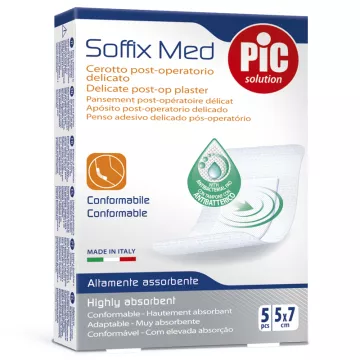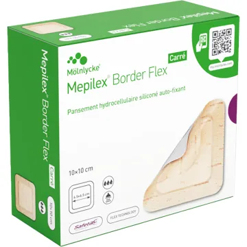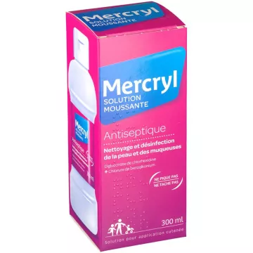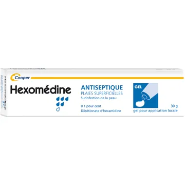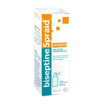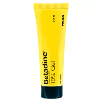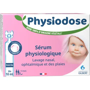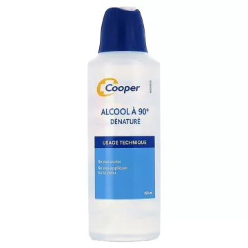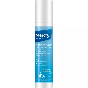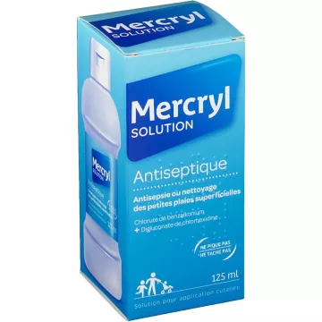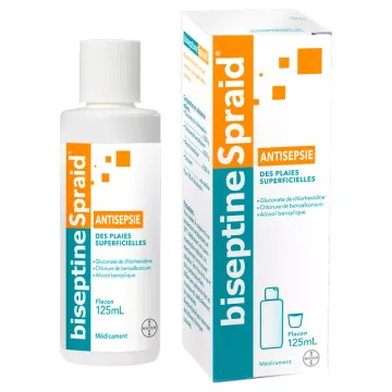
Wound care always begins with thorough mechanical cleansing: rinse the wound thoroughly with sterile saline solution or clean running water for at least 5 minutes to remove foreign objects, debris, and bacteria. The antiseptic is applied after mechanical cleansing—never instead of it. For cuts and superficial wounds, chlorhexidine (0.5% solution) is the antiseptic recommended by the HAS: it is a broad-spectrum bactericide, well-tolerated, and non-cytotoxic at standard concentrations. Povidone-iodine (10%) is effective for wounds at high risk of infection but may slow healing at high concentrations. Hexamidine (0.1% gel) is an alcohol-free antiseptic that is well tolerated on painful wounds and sensitive skin.
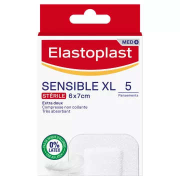
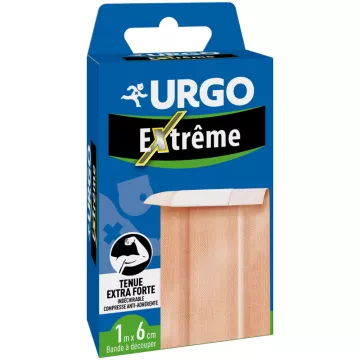
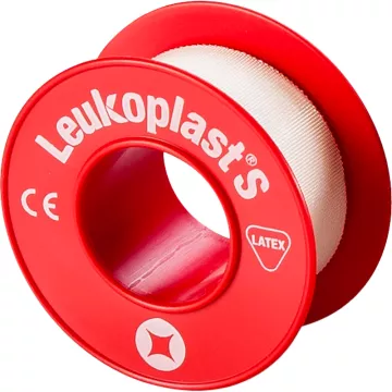
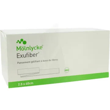
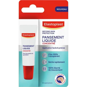
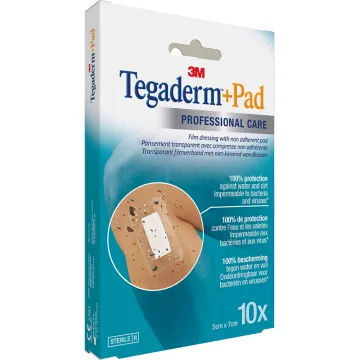
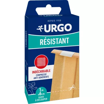
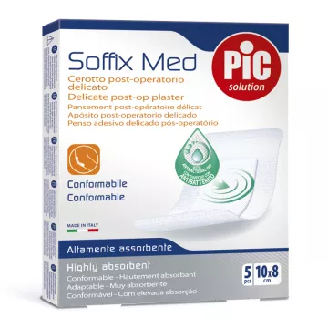
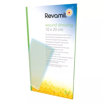
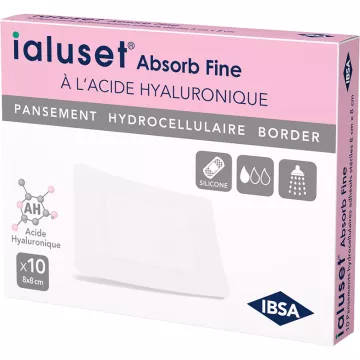
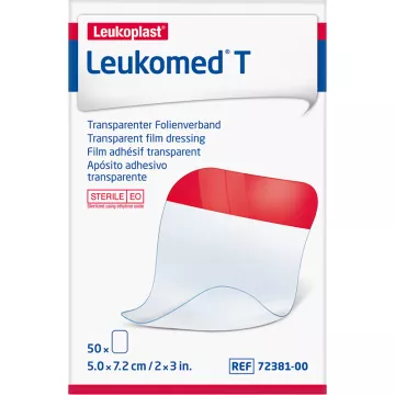
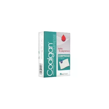
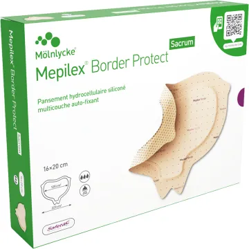
The choice of dressing depends on the type and stage of the wound:
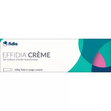
Hyaluronic acid (HA) is a glycosaminoglycan naturally present in the dermis that plays a key role in the three phases of wound healing (inflammation, proliferation, and remodeling). High-molecular-weight HA creams form an occlusive film that locks in moisture, promotes fibroblast migration, and accelerates epidermal closure. Low-molecular-weight HA penetrates the dermis and stimulates collagen synthesis—particularly useful for deep wounds and recent scars. Apply only to clean, disinfected wounds—never to infected wounds.
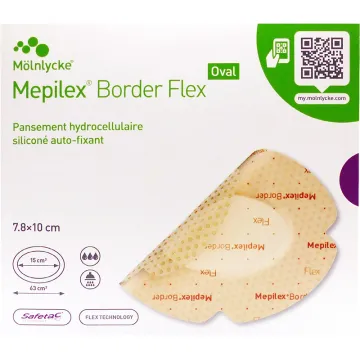
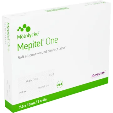
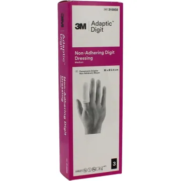
First-degree and superficial second-degree burns (blisters covering less than 1% of the body surface area) can be treated at home. First step: Immediately cool the area under cold running water (15–25°C) for at least 15 minutes—never use ice. After cooling, apply a hydrocolloid or hydrocellular dressing to maintain moisture and protect against contamination. Never puncture blisters. Abrasions should be treated like any other wound: clean with saline solution or an antiseptic, then apply a sterile compress held in place with a crepe bandage. Non-adherent gauze (oily tulle or a silicone interface) is recommended for very painful wounds.
An infected wound is characterized by local signs: extensive redness, warmth, swelling, pus, a foul odor, and yellow crusts extending beyond the wound edges. General signs (fever, a red streak extending toward the lymph node—lymphangitis) require urgent medical attention. Deep wounds, bites (from humans or animals), wounds contaminated with dirt, or wounds in diabetic or immunocompromised patients must always be medically evaluated—the risk of tetanus must be systematically assessed. A wound that does not heal within 3 weeks despite appropriate care requires a dermatological evaluation.
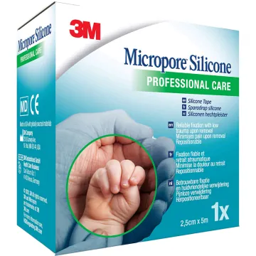
Scar prevention begins as soon as the wound closes. Sun protection (SPF 50+) is essential for 12 to 18 months—UV rays stimulate melanogenesis and cause long-lasting post-inflammatory hyperpigmentation. Silicone gels and patches (12 hours a day for 3 to 6 months) are the validated gold-standard treatments for reducing hypertrophy and improving texture. Scar massage (5 minutes a day with a rich cream) promotes collagen maturation. For keloids or stubborn hypertrophic scars, a dermatology consultation is necessary. Sunburn on a recent scar is particularly damaging and must be avoided during the first year.