What is a hematoma, and how does it form?
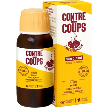
A hematoma is a collection of clotted or liquid blood that forms outside blood vessels, in soft tissues or an organ cavity, following the rupture of one or more blood vessels. Unlike a bruise (superficial bleeding) or a blue mark, a hematoma is palpable—it forms a lump or swelling under the skin. Some hematomas are benign and resolve on their own, while others constitute absolute medical emergencies. Treatment for ecchymoses and contusions is available in the “Bumps and Bruises” section of the store.
- Types of hematomas based on location:
- Subcutaneous: the most common—a palpable, painful, bluish mass under the skin—resulting from direct impact, venipuncture, or anticoagulant use
- Intramuscular: following significant trauma or an IM injection—may compress nerve structures—risk of compartment syndrome (surgical emergency)
- Intracranial (subdural, epidural): neurosurgical emergency—following head trauma (even minor in patients on anticoagulants)—warning signs: severe headache, progressive drowsiness, vomiting, neurological deficit → call 15 immediately
- Postoperative: a common complication following surgery—especially in cases of coagulation disorders or in patients on anticoagulants—surgical drainage if the hematoma is large or compressive
- Risk factors: anticoagulants (warfarin, DOACs, LMWH) — antiplatelet agents (aspirin, clopidogrel) — coagulation disorders (hemophilia, von Willebrand disease) — vitamin C or K deficiency — capillary fragility — long-term corticosteroid therapy
- Distinguishing between a hematoma, ecchymosis, and a bruise: hematoma = palpable mass — ecchymosis = flat, non-palpable discoloration — see the dedicated pages for specific care
Warning signs and management of a hematoma
- Benign hematoma (conservative management): subcutaneous or superficial intramuscular hematoma following identifiable trauma — stable size — moderate pain that is improving — color changing to green then yellow-brown (normal resolution within 7 to 14 days)
- Call 15 immediately if: head trauma accompanied by headaches, drowsiness, vomiting, confusion, or neurological deficits (suspected intracranial hematoma)—expansive hematoma of a limb (rapid increase in volume) with severe pain and paresthesias (compartment syndrome) — abdominal hematoma with hemodynamic instability (pallor, hypotension, tachycardia)
- See a doctor if: spontaneous hematoma without identifiable trauma (suspected coagulation disorder) — large hematoma while on anticoagulants — secondary infection (warmth, redness, fever) — periorbital hematoma following a head injury (“raccoon eyes” — skull base fracture)
Natural treatments and care to speed up the resolution of a hematoma
- Cold/hot protocol: cold (0–48 h) — wrapped ice pack for 15 to 20 min, 3 to 4 times a day—vasoconstriction limits spread—never apply directly to the skin—gentle heat (after 48 hours)—warm compress—vasodilation promotes the resorption of clotted blood
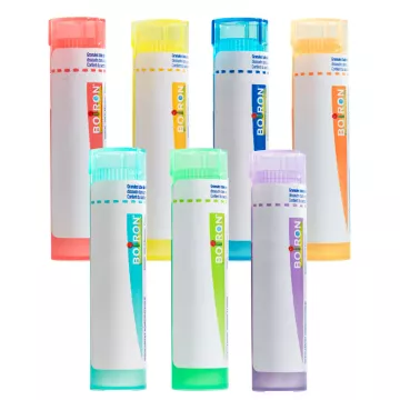
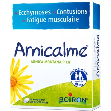
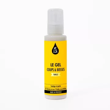
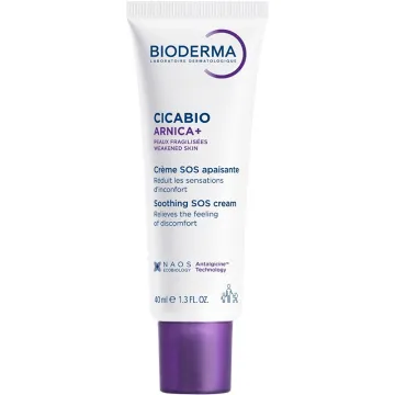
- Arnica montana: anti-inflammatory and anti-edematous helenine and helicoside — reduces swelling and accelerates the resolution of the hematoma — topical gel or cream 2 to 3 times daily — never apply to broken skin or open wounds — homeopathic use (Arnica 5 CH, 5 granules 3 times daily)
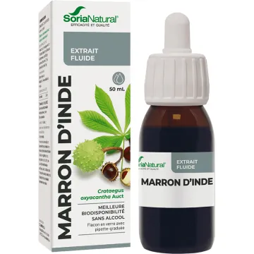
- Escin (horse chestnut): anti-edematous and vasoconstrictive — reduces capillary permeability and accelerates resorption — as a topical gel or in capsules — Contraindicated with anticoagulants (consult a doctor)
- Vitamin C + bioflavonoids: strengthens capillary walls — reduces vascular fragility — prevents recurrent bruising of nutritional origin