
Health information prepared under the supervisionof Arnaud, Ph.D. in Pharmacy. Sources: Haute Autorité de Santé (HAS), Société Française de Dermatologie (SFD), Inserm, International Agency for Research on Cancer (IARC). This article is not a substitute for a dermatological consultation. If you notice any suspicious lesions, consult a dermatologist.
Actinic keratosis (AK), also known as solar keratosis, is a precancerous skin lesion caused by chronic exposure to ultraviolet (UV) rays— whether natural (sunlight) or artificial (tanning beds, classified as Group 1 carcinogens by the IARC). It develops on areas of the skin chronically exposed to sunlight: the face (forehead, nose, cheekbones, temples), ears, lips (actinic cheilitis), scalp in people with alopecia, backs of the hands, forearms, and décolletage.
Actinic keratosis is part of a dermatological continuum that, if left untreated, can progress to cutaneous squamous cell carcinoma. The individual risk of transformation from a single lesion is low, but when accumulated over a large number of lesions and several years, it becomes significant. The concept of a “field of carcinogenesis” is central: the area surrounding visible lesions is also exposed and harbors subclinical cellular abnormalities.
Actinic keratosis lesions have characteristic clinical features:
Warning signs requiring immediatedermatological consultation: a lesion that thickens rapidly, bleeds at the slightest trauma, ulcerates, becomes spontaneously painful, or exceeds 1 cm in size. These signs may indicate a potential transformation into squamous cell carcinoma.
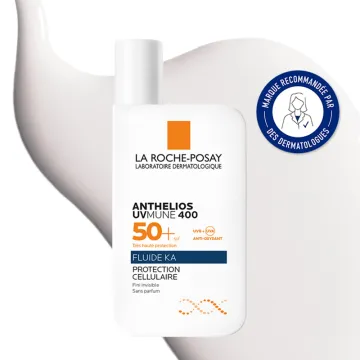
Prevention relies on rigorous sun protection throughout one’s life:
Several studies (notably the ONTRAC trial published in the New England Journal of Medicine in 2015, Chen et al.) have documented a moderate protective effect of oral nicotinamide (vitamin B3) at a dose of 500 mg twice daily in high-risk patients, with a reduction of approximately 23% in actinic keratoses. This approach should be discussed with a dermatologist before starting any long-term supplementation.
The choice of treatment depends on the number of lesions, their location, their thickness, and the clinical context. All treatments are prescribed and administered by a dermatologist:
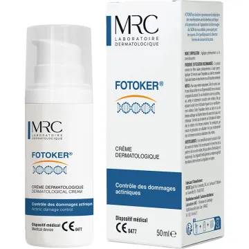
No cosmetic treatment or dietary supplement can replace these medical interventions. However, dermo-cosmetic products (moisturizers, reparative balms, ceramides, after-sun treatments) may support post-treatment skin recovery, as advised by a dermatologist.
The diagnosis is made by a dermatologist based on a clinical examination, including visual inspection and palpation of the lesions, as well as a comprehensive examination of the skin. The examination may be supplemented by:
The dermatologist also assesses the area at risk for cancer and other skin lesions (carcinomas, nevi requiring monitoring). In cases of uncertainty regarding other lesions, advanced photoaging of the skin may accompany the keratoses in the same areas.
Yes, actinic keratosis is considered a precursor to cutaneous squamous cell carcinoma. According to available data (HAS, SFD), the risk of transformation per isolated lesion per year is low (estimates ranging from 0.025% to 16% depending on the study, most often around 1% per lesion per year), but this risk becomes significant when the lesions are numerous, long-standing, or located within an extensive field of precancerous changes.
Cutaneous squamous cell carcinoma, when detected early, generally has a good prognosis. Regular dermatological follow-up is therefore key to optimal management. Any lesion that thickens, bleeds, ulcerates, or becomes painful should be evaluated promptly.
With appropriate dermatological care and rigorous sun protection, the prognosis is favorable in the vast majority of cases. Current treatments allow for the effective eradication of lesions, and prevention of recurrence relies on:
People with actinic keratoses have an increased risk of developing other skin cancers (basal cell carcinomas, melanomas); therefore, dermatological follow-up includes a complete examination of the skin.
Actinic keratosis is a very common condition, the prevalence of which increases with age and latitude (high prevalence in countries with intense sunlight). In Europe, several studies (Werner 2013, Heerfordt 2017) report a prevalence of approximately 15 to 25% among adults over 60 with fair skin types, with higher rates in Australia and Mediterranean countries.
The increase in outdoor activities, summer tourism, and the historical use of tanning beds also explains its occurrence in younger adults (ages 40–50), particularly among those who experienced repeated sunburns during childhood and adolescence.
Several individual risk factors have been identified:
Some lesions may regress spontaneously, but they tend to recur at the same site. Studies report varying rates of spontaneous regression (up to 25% at one year for certain cohorts), but an equal or greater proportion of lesions persist or reappear. Lack of treatment exposes patients to the risk of progression and transformation.
It is therefore not recommended to wait for “spontaneous resolution”: any suspicious lesion should be evaluated by a dermatologist, who will decide on treatment or simply monitoring depending on the circumstances.
The differential diagnosis includes several skin lesions that may resemble actinic keratosis:
The dermatologist distinguishes these lesions through clinical examination, dermoscopy, and, if necessary, a biopsy. No remote self-assessment can replace this examination.
Living with actinic keratosis can cause significant anxiety, related to the visible appearance of the lesions on the face and the fear that they may develop into cancer. The treatments themselves (redness, scabbing, and a burning sensation lasting 1 to 3 weeks, depending on the technique) can affect self-image and social life.
Here are some suggestions for managing these issues:
Regular follow-up and strict sun protection help restore confidence in managing the condition over the long term.
This article is for informational purposes only. If you notice a suspicious skin lesion (rough, persistent, thickening, bleeding, or ulcerating), make an appointment with a dermatologist. Early detection leads to simpler treatment and a better prognosis.