What is an oral lesion, and what are the main types?
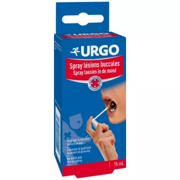
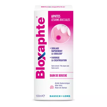
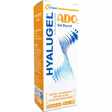
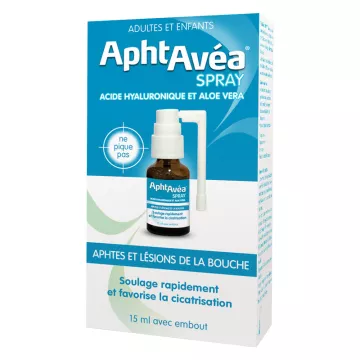
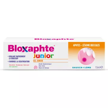
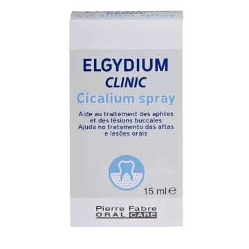
The term “oral lesions” refers to any abnormal change in the tissues of the oral cavity—gums, tongue, palate, inner cheeks, lips, and floor of the mouth. These lesions range from a common canker sore that heals on its own within a week to a precancerous lesion that requires a biopsy. The main challenge is to quickly distinguish a benign lesion from a suspicious one—and the two-week rule applies universally: any oral lesion that does not heal spontaneously within 14 days should be examined by a doctor or dentist.Oral hygiene care and canker sore treatments available in the store can help manage common benign lesions.
- Ulcerative lesions: loss of mucosal tissue—canker sores (round, painful ulcers with a yellow center bordered by red, healing in 7–14 days) are the most common form—see the page dedicated to canker sores —herpetic ulcers are more superficial and clustered—traumatic ulcers (caused by biting or ill-fitting dentures) are often unilateral and localized at the site of friction
- White lesions: leukoplakia (white patch that does not disappear when scraped → biopsy required, as 5–17% may become malignant) — oral candidiasis / thrush (white patches that disappear when scraped → antifungal treatment — see candidiasis) — oral lichen planus (symmetrical, reticulated white streaks — an autoimmune condition, noncontagious)
- Red (erythematous) lesions: erythroplasia (persistent velvety red patch → high risk of malignancy, urgent biopsy) — stomatitis (diffuse inflammation — see stomatitis) — gingivitis (redness + bleeding gums) — contact erythema (local allergen)
- Vesicular and bullous lesions: herpes simplex (cold sores —clustered vesicles on an erythematous base) — shingles (VZV — unilateral vesicles strictly confined to a nerve distribution) — pemphigus vulgaris (fragile blisters — serious autoimmune disease, urgent dermatological consultation)
- Pigmented and neoplastic lesions: benign oral melanosis (flat, stable brown spot) — mucosal melanoma (rare but serious—rapid growth, irregular borders) — traumatic fibromas (firm, painless nodules caused by repeated friction) — papilloma (HPV—cauliflower-like growth)
What deficiencies can cause recurrent oral lesions?
- Iron deficiency: Iron deficiency is the leading nutritional cause of recurrent oral lesions—the oral mucosa renews itself rapidly, and iron is essential for this process—signs: recurrent canker sores + atrophic glossitis (red, smooth, non-papillated tongue) + cracks at the corners of the mouth + fatigue + pallor — a ferritin test is required before any prolonged symptomatic treatment
- Vitamin B12 deficiency: Vitamin B12 is necessary for the rapid division of oral epithelial cells—a deficiency causes glossitis (red, painful, smooth tongue) and mucosal ulcers—suspect this in vegans, the elderly, and patients taking PPIs or metformin
- Folic acid (B9) deficiency: Folic acid, along with vitamin B12, is involved in DNA synthesis in mucosal cells—a deficiency causes symptoms similar to those of vitamin B12 deficiency (glossitis, stomatitis, ulcerations)—particularly common in women taking oral contraceptives and chronic alcoholics
- Zinc deficiency: Zinc is a cofactor for local mucosal immunity and epithelial repair—a deficiency slows the healing of lesions, promotes recurrent ulcers, and may be accompanied by a loss of taste (ageusia)—zinc combined with vitamin C accelerates mucosal healing
- Undiagnosed celiac disease: recurrent oral aphthous ulcers are sometimes a telltale sign of gluten-induced enteropathy—in cases of multiple, very frequent aphthous ulcers that are resistant to standard treatments, plus multiple deficiencies (B12, iron, folate) → request a celiac serology test (IgA anti-transglutaminase antibodies) from your doctor
How can you distinguish a benign lesion from a suspicious one?
- Reassuring features of a benign lesion: recent onset following an identifiable trigger (bite, hard food, stress, viral infection) — size < 1 cm — sharp, regular edges — pain present (paradoxically, malignant lesions are often less painful) — spontaneous healing within 14 days
- Warning signs requiring prompt medical attention: a lesion that persists for more than 2 weeks without an obvious cause — a white or red patch that does not disappear when scraped — irregular, indurated (hard to the touch) edges — a mixture of red and white (erythroleucoplastic lesion) — rapid growth — painless (early-stage oral cancers are often barely painful or painless) — associated cervical lymphadenopathy (hard lymph node under the jaw)
- Risk factors for oral cancer: tobacco (cigarettes, pipes, cigars, chewing tobacco—the primary risk factor) + alcohol (synergistic effect with tobacco—the risk is 15 times higher among smokers who also drink) + HPV 16 and 18 (on the rise, particularly for oropharyngeal cancers) + sun exposure (lower lip) + chronic poor oral hygiene — anyone with these risk factors should have regular oral exams at the dentist
- Personal mirror exam: a monthly self-examination of the mouth (lips, tongue, cheeks, palate, gums, floor of the mouth) helps detect any unusual changes quickly—the dentist performs this examination routinely during appointments—report any changes noticed since your last visit
How can you relieve mild oral lesions on a daily basis?
- Saltwater or baking soda mouthwashes: 1 teaspoon of salt or baking soda in a glass of lukewarm water—rinse 3 to 4 times a day—a mild local anti-inflammatory that cleans the lesions and alkalizes the oral pH (creating a hostile environment for bacteria)—provides rapid relief without irritating the mucous membrane—an alternative to alcohol-based antiseptic mouthwashes that can cause irritation
- Local anesthetic gels: 2% lidocaine topical gel — apply before meals to allow for pain-free eating — fast-acting (2–3 min) but short-lasting (30–45 min) — available over the counter at pharmacies — do not exceed the recommended doses (risk of systemic toxicity at very high doses)
- Manuka honey: documented antibacterial properties (methylglyoxal — MGO ≥ 250) — reduces inflammation and local pain in ulcers — apply directly to the lesion 3 to 4 times a day — suitable for canker sores, mild herpes lesions, and minor stomatitis
- Appropriate diet during the acute phase: avoid acidic foods (citrus fruits, tomatoes, vinegar), spicy foods, and foods that are very hot or very cold — opt for soft and cool textures (yogurt, fruit compote, lukewarm purees)—cut food into small pieces to reduce friction on the lesions—stay well-hydrated to keep saliva flowing (the mucous membranes’ first natural defense)
- When to seek medical attention: a lesion that does not heal within 14 days—severe pain despite pain relievers—difficulty swallowing, breathing, or opening the mouth—associated fever—a lesion in an immunocompromised patient (HIV, chemotherapy, long-term corticosteroid use)—uncertainty about the nature of the sore—for toothaches and associated dental abscesses, emergency dental care is required