What is a pressure ulcer?
A pressure ulcer is a localized ischemic lesion of the skin and underlying tissues caused by prolonged compression of soft tissues between a bony prominence and a pressure surface. According to the international NPUAP-EPUAP-PPPIA definitions (2019), it may involve pressure, shear, and friction on skin made vulnerable by immobility, malnutrition, moisture, advanced age, or illness.
Typical sites are located on the pressure points of bony prominences:
- Sacrum, ischial tuberosities, trochanters (while sitting or lying down).
- Heels, malleoli, outer edges of the feet.
- Elbows, shoulders, occiput (during prolonged supine positioning).
- Knees, inner sides when lying on one’s side.
- Edge of the nose (nasogastric tube), ears (oxygen goggles, mask), pressure points from casts or splints.
More rarely, “iatrogenic pressure ulcers” develop at unintended pressure points caused by medical devices (tubes, tubing, catheters, belts). See also “Wounds” for an overview of skin care.
How can pressure ulcers be prevented?
Prevention is the cornerstone of care:
- Systematic risk assessment using a validated scale (Braden—especially in France—or Norton) upon admission or when a patient’s dependency status worsens, followed by regular reassessment.
- Mobilization and repositioning every 2 to 3 hours (day and night), alternating pressure points (supine, right and left semi-lateral, heel relief). A schedule is helpful for caregivers.
- Relieve pressure on at-risk pressure points through proper positioning (using pillows and positioning cushions), with particular attention to the heels (which should be elevated with a cushion under the calf to completely relieve pressure).
- Appropriate therapeutic supports: memory foam mattresses, static or dynamic air mattresses, alternating pressure mattresses—selection based on risk level. Specialized seat cushions for individuals in wheelchairs.
- Daily skin care: gentle cleansing with lukewarm water and a mild soap substitute, drying by gently patting, moisturizing with a well-tolerated emollient, paying attention toskin folds and macerated areas. No vigorous massage of bony prominences (contraindicated—see below). Opt for gentle stroking with an emollient.
- Maintaining dry skin: appropriate changes of clothing in cases of incontinence, skin protection (protective films, zinc oxide barrier creams), and management of sweating.
- Earlynutritional assessment and support (see dedicated section).
- Regular oralhydration.
- Active mobilization as soon as possible (physical therapy, transferring to a chair, walking).
- Informing the patient and their family and friends: prevention is the responsibility of the multidisciplinary team and daily caregivers.
What are the stages of pressure ulcers?
The international NPUAP-EPUAP-PPPIA classification distinguishes several categories:
- Stage 1: intact skin with non-blanching erythema (redness persists when pressure is applied with a finger to the area). First warning sign—it’s time to take active measures.
- Stage 2: Partial loss of the dermis appearing as a superficial, open, shallow ulcer with a red-pink base and no black necrosis. May also take the form of an intact or ruptured phlycten (serous blister).
- Stage 3: Total skin loss exposing the subcutaneous fat. Possible detachment beneath the edges; the base is filled with granulation tissue. Muscle, bone, or tendons are not exposed.
- Stage 4: Total tissue loss with exposure of bone, tendon, or muscle. Frequent presence of black or yellow necrosis (slough), fistulas, and deep detachments.
- Deep tissue injury: a purplish or dark brown area on intact skin, or a hemorrhagic phlycten, indicating underlying deep tissue damage. May rapidly progress to Stage 3–4.
- Unclassifiable pressure ulcer: total tissue loss whose depth cannot be assessed because the base is entirely covered by black or yellow necrosis. The stage will be determined only after debridement.
What is the treatment for pressure ulcers?
Management is tailored to the stage and individualized for each person, based on a doctor’s prescription and in coordination with the nurse:
- Stage 1: Immediate relief of pressure on the affected area, more frequent repositioning, close monitoring, emollients, and very gentle massage, along with appropriate therapeutic support.
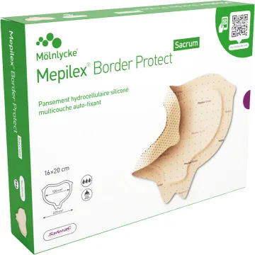
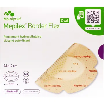
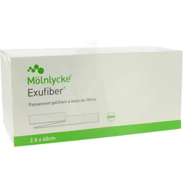
- Stage 2: Modern moist dressings (thin hydrocolloids, polyurethane films, non-adherent interface dressings, hydrofibers for moderate exudate). Mechanical protection. Care of the skin surrounding the lesion.
- Stages 3–4:
- Debridement of devitalized tissue: autolytic (hydrogels, moist dressings), mechanical (moist compresses, water jet), enzymatic, surgical (curette, scalpel), and larval therapy in specialized settings for resistant sloughy wounds.
- Dressings tailored to the phase: hydrogels (debridement), alginates (exudates), hydrofibers, absorbent foams, and dressings containing silver or certified medical-grade honey in cases of colonization or infection.
- Negative pressure wound therapy (NPWT/VAC): useful for certain deep, cavitary, or exudative wounds, in a hospital or home health care setting.
- Pain relievers appropriate for the intensity of the pain (pressure ulcers are often painful, particularly during dressing changes).
- Systemic antibiotic therapy as prescribed in cases of local or systemic signs of infection. No routine topical antibiotic therapy. See “Infected Wounds.”
- Reconstructive surgery (grafts, flaps) performed in a specialized setting for deep, refractory pressure ulcers, following preparation of the wound bed.
- Hydrogen peroxide is no longer used routinely (it is cytotoxic to granulation tissue and delays healing).
- Targetedantisepsis (aqueous chlorhexidine, povidone-iodine with rinsing) when specifically indicated, without prolonged use.
- See also “Wound Healing” for general principles of wound care.
What complications can arise from pressure ulcers?
Complications from pressure ulcers can be severe:
- Local infection: colonization followed by bacterial infection (often polymicrobial, including *Staphylococcus aureus*, streptococci, enterobacteria, and anaerobes).
- Cellulitis, dermo-hypodermitis: spread of the infection to the surrounding soft tissues.
- Osteitis, osteomyelitis: bone infection, particularly dangerous in sacral and trochanteric pressure ulcers.
- Bacteremia, sepsis: entry of bacteria into the bloodstream, a life-threatening emergency in frail individuals.
- Fistulas between the pressure ulcer and deep structures (bladder, rectum, joint).
- Worsening of malnutrition due to protein loss associated with the chronic wound.
- Chronicanemia, hypoalbuminemia.
- Psychological impact: chronic pain, dependence, impaired self-image, and a sense of being a burden to loved ones.
- Increased mortality in frail older adults, particularly in cases of deep, infected pressure ulcers.
Skin weakened by age or medical treatments (corticosteroids, anticoagulants) increases the risk of complications.
How are pressure ulcers diagnosed?
The diagnosis is primarily clinical:
- A thorough examination of the skin, with particular attention to pressure points, including areas under medical devices and the heels.
- Staging according to NPUAP-EPUAP-PPPIA criteria.
- Measurement of the wound (length, width, depth), description of the wound bed (granulation tissue, fibrin, necrosis), wound margins, exudate (volume, appearance, odor), and the skin surrounding the lesion.
- Pain assessment (appropriate scales: VAS, EN, behavioral scales for non-communicative patients).
- Risk assessment using the Braden scale (6 items: sensory perception, moisture, activity, mobility, nutrition, friction and shear; score <18 = at risk, <12 = high risk) or the Norton scale.
- Laboratory workup as indicated by the clinical context: CBC, CRP, serum albumin, prealbumin, blood glucose, renal function.
- Imaging (ultrasound, CT scan, MRI) if deep extension, fistula, or osteitis is suspected.
- Bacteriological culture if signs of infection are present (not routine).
- Dated photographs for monitoring progression.
What are the risk factors associated with pressure ulcers?
Factors to consider when identifying at-risk individuals:
- Immobility or reduced mobility: prolonged bed rest, wheelchair use, paralysis, postoperative conditions, cognitive impairments that limit the perception of discomfort.
- Malnutrition: unintentional weight loss, low BMI, low serum albumin, insufficient protein intake.
- Excessive moisture: urinary and/or fecal incontinence, sweating, wound exudate.
- Pressure and shear forces: prolonged sitting, sliding in bed, handling without appropriate support.
- Sensory perception disorders: neuropathies, cognitive impairments, sedation, anesthesia.
- Circulatory disorders: arterial disease, venous insufficiency, anemia, tissue hypoxia.
- Chronic diseases: diabetes, renal failure, cancer, COPD, dementia, Parkinson’s disease.
- Medications: long-term corticosteroids, immunosuppressants, anticoagulants (triggering hematoma).
- Advanced age: geriatric skin is thinner, more fragile, less elastic, and less well-vascularized.
- History of pressure ulcers.
- Medical devices: catheters, masks, tubes, splints, casts.
Can pressure ulcers be treated at home?
Yes, in many situations, home care is possible and even desirable, provided it is properly organized:
- Coordination by the primary care physician and the assigned independent nurse.
- Regular nursing care, with the frequency tailored to the stage of the ulcer and the amount of exudate.
- Provision of appropriate equipment: therapeutic mattresses/cushions, modern dressings (by prescription), and care products.
- Home hospitalization (HAD) for complex, exudative, or deep wounds, or those requiring more technical care (total parenteral nutrition, IV antibiotic therapy).
- City-hospital networks, mobile wound and healing teams, and telemedicine between the local registered nurse and an expert consultant.
- Training and support for family caregivers regarding repositioning, skin care, and monitoring of new at-risk areas.
- Social support (assistance with personal hygiene, meal delivery, in-home physical therapy).
- Nutritional assessment by the attending physician or a dietitian, with oral nutritional supplements if necessary.
- Hospitalization if the condition worsens, there is a deep infection, complications arise, or the patient’s overall condition deteriorates.
What role does nutrition play in preventing pressure ulcers?
Nutrition is a major determinant of prevention and wound healing:
- Appropriate energy intake: 30 to 35 kcal/kg/day as a general rule, more in cases of severe wounds or hypercatabolism.
- High protein intake: 1.2 to 1.5 g/kg/day for prevention in at-risk individuals, up to 1.5 to 2 g/kg/day in the presence of pressure ulcers, unless there is renal insufficiency (ESPEN/HAS recommendations).
- Regularhydration: approximately 30 ml/kg/day.
- Vitamin C: an essential cofactor for collagen synthesis. Vary dietary sources; supplement if a biological deficiency is present.
- Zinc: involved in wound healing and immunity. Supplementation if a documented biological deficiency exists.
- Vitamin A, vitamin D, iron, selenium, B vitamins: overall balance is important.
- Oral nutritional supplements (ONS) by prescription if malnutrition is present (weight loss >5% in 1 month or >10% in 6 months, low BMI, serum albumin <35 g/L).
- Enteral nutrition in certain severe cases, as prescribed.
- Follow-up by a dietitian, particularly in geriatrics and chronic wound care.
- Regular assessment (MNA, weight monitoring, laboratory tests).
Are pressure ulcers always preventable?
Prevention is highly effective when implemented early and comprehensively, but certain situations partially defy this goal:
- End-of-life conditions: changes in cutaneous perfusion and tissue fragility can make a pressure ulcer unavoidable despite optimal care. This is sometimes referred to as a “terminal pressure ulcer” or “Kennedy Terminal Ulcer,” which has a different pathophysiological mechanism.
- Situations of major hemodynamic instability (resuscitation, shock) where the priority is on vital functions.
- Respiratory distress requiring restricted positioning (prone position in ARDS, invasive ventilation).
- Essential medical devices that exert prolonged pressure on the skin.
- Refusal by the patient or their caregivers to follow part of the prevention protocol.
In the vast majority of cases, early risk assessment, appropriate prevention, team vigilance, and proper nutrition can prevent the development of pressure ulcers or limit their severity. The occurrence of a pressure ulcer, especially in geriatric care, should prompt a review of the organization of care rather than a search for blame. The role of caregivers, home health aides, physical therapists, and mobile wound care teams is central to this multidisciplinary approach.