What is a varicose ulcer, and how does it develop?
A varicose ulcer (or venous ulcer) is a chronic, non-healing open wound resulting from severe chronic venous hypertension—it corresponds to stage C6 (active ulcer) or C5 (healed ulcer) stage of the CEAP classification. It is the most serious complication ofchronic venous insufficiency. Any leg wound that does not heal within 2 to 4 weeks should be evaluated by a doctor. Venous care products are available in the “heavy legs ” and “venous circulation ” sections of the store.
- Pathophysiological mechanism: varicose veins and incompetent valves → chronic venous hypertension → chronic microinflammation → extravasation of fibrin and red blood cells → pericapillary fibrin deposition (barrier to O₂ diffusion) → tissue hypoxia → skin necrosis → ulceration
- Typical location and appearance: inner side of the ankle (Gaiter’s area) — broad base, irregular edges, clean pink or fibrinous center — surrounded by ochre dermatitis (ferruginous hyperpigmentation) and lipodermatosclerosis (indurated, brownish skin) — often not very painful (unlike an arterial ulcer)
- Risk factors: Advanced, untreated chronic venous insufficiency (CEAP ≥ C3) — history of deep vein thrombosis (post-thrombotic syndrome) — prolonged immobilization — obesity — smoking — diabetes (a factor that impairs healing)
Differential diagnosis and complications
- Venous ulcer vs. arterial ulcer vs. diabetic ulcer: Arterial ulcers (AOMI) are more painful, have a punch-hole appearance, and are located distally (toes, top of the foot) — Diabetic ulcers are predominantly plantar (pressure area) — the differential diagnosis is medical and relies on arterial and venous Doppler ultrasound — NEVER apply firm compression to an arterial ulcer (worsens ischemia)
- Complications of varicose ulcers: bacterial infection (erysipelas, cellulitis—fever + extensive redness + pain → antibiotics)—contact dermatitis from topical treatments—rare malignancy (Marjolin’s squamous cell carcinoma on a chronic ulcer)—secondary amyloidosis—significant impairment of quality of life
- See a doctor or vascular specialist immediately if: any leg wound persisting for > 2 to 4 weeks — signs of infection (fever, extensive redness, odor) — ulcer enlarging despite treatment — suspected arterial ulcer (pain when lying down, cold foot)
Treatment of Varicose Ulcers and Venous Support
The treatment of varicose ulcers relies on specialized medical care—a home health nurse for wound care, and an angiologist for vascular assessment and compression therapy. These complementary measures support a healthy venous lifestyle—they do not replace professional wound care.
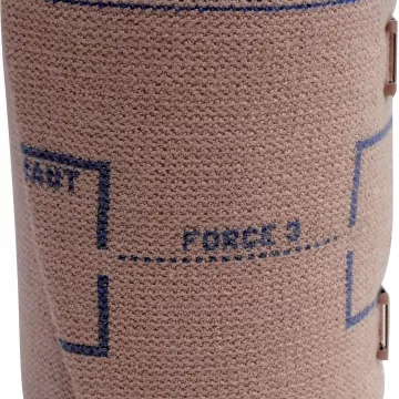
- Standard therapeutic compression: multilayer bandages (sterile + absorbent + 35–40 mmHg compression) applied by a nurse—Class 3 or 4 compression stockings after healing to prevent recurrence — NEVER use high-compression bandages without a prior vascular assessment (risk of ischemia)
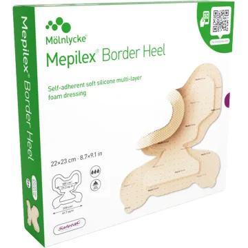
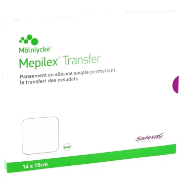
- Local wound care: gentle cleansing with saline solution — dressings appropriate for the stageof healing (hydrocolloid on clean wounds, hydrofiber if exudate is present, charcoal if infection is present) — as prescribed by a physician or nurse
- Bioflavonoids (diosmin, hesperidin): reduce venous hypertension and capillary permeability — accelerate the healing of venous ulcers in several clinical trials — to be taken as a supplement to medical treatment
- Aescin and red vine extract: support venous tone and reduce peri-ulcer edema — available in capsules — contraindicated with anticoagulants
- Prevention of recurrence: long-term use of Class 2–3 compression stockings — regular physical activity (30-minute walk daily) — surgical treatment of underlying varicose veins (sclerotherapy, stripping) — maintaining a healthy weight — regular follow-up with a nurse or dermatologist